 Jun, 3 2026
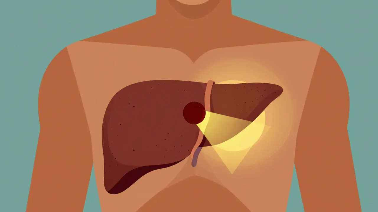
Imagine finding a small tumor in your liver before it causes any pain or visible symptoms. For people with cirrhosis is a late stage of scarring (fibrosis) of the liver caused by many forms of liver diseases and conditions, this scenario isn't just hopeful-it's the standard goal of modern medicine. Hepatocellular carcinoma is the most common type of primary liver cancer, accounting for 75-85% of all liver cancer cases globally. Without regular checks, this disease often hides until it’s too late for curative options. But with proper surveillance, survival rates jump from roughly 10-20% to 50-70%. The difference between catching it early and missing it entirely can mean the difference between life-saving surgery and palliative care.
Why Cirrhosis Is the Main Risk Factor
You don’t need to be an expert to know that a damaged liver is vulnerable. Over 80% of hepatocellular carcinoma cases occur in patients who already have advanced fibrosis or cirrhosis. This makes cirrhosis the single biggest predictor for developing liver cancer. Whether your liver damage comes from chronic hepatitis B, hepatitis C, alcohol use, or non-alcoholic fatty liver disease (NAFLD), the scar tissue creates an environment where cancer cells can thrive.
The risk isn't equal for everyone. If you have hepatitis B, your annual risk of developing HCC sits between 5% and 8%. For those with NAFLD, especially after achieving sustained virologic response (SVR) for other conditions, the risk is lower, around 1% to 3%. Understanding your specific baseline helps you and your doctor decide how aggressively to monitor your health.
How Surveillance Works: The Standard Protocol
Surveillance isn't about guessing; it's about following a strict schedule designed to catch tumors when they are still tiny-usually under 2 centimeters. The gold standard, recommended by the American Association for the Study of Liver Diseases (AASLD) is a professional organization dedicated to advancing research and clinical practice in liver diseases and other major bodies, involves two main tools:
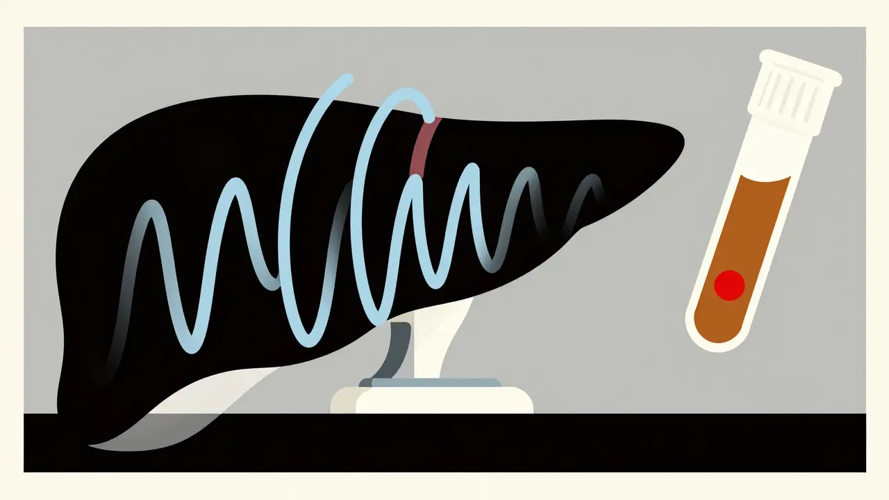
- Ultrasound Imaging: You get an abdominal ultrasound every six months. Why six months? Because data shows HCC tumors typically grow by 1-2 cm in that timeframe. Catching them within this window keeps them treatable.
- Alpha-Fetoprotein (AFP) Blood Test: This is often added to the mix. While not perfect on its own, an AFP level above 20 ng/mL acts as a red flag that triggers immediate further investigation.
If either test shows something unusual-a mass larger than 1 cm on the ultrasound or elevated AFP-the next step is clear. You undergo multiphase contrast imaging, such as a CT scan or MRI. These advanced scans have a sensitivity rate of 80-90% for characterizing lesions, helping doctors confirm if it’s cancer without needing an invasive biopsy immediately.
New Directions: Risk-Based Surveillance
Not all cirrhosis is created equal. In April 2023, the European Association for the Study of the Liver (EASL) published Clinical Practice Guidelines and Policy Statements for liver cancer management introduced a smarter approach: risk-based surveillance. Instead of scanning everyone equally, they categorize patients into three tiers based on their annual HCC risk:
| Risk Tier | Annual HCC Risk | Recommended Action |
|---|---|---|
| High-Risk | >2.5% | 6-month Ultrasound or potentially MRI |
| Medium-Risk | 1.5-2.5% | Standard 6-month Ultrasound |
| Low-Risk | <1.5% | Potentially forgo routine surveillance |
This model could reduce unnecessary tests by 20-30%, saving money and reducing patient anxiety. Tools like the aMAP score (which looks at age, gender, albumin, bilirubin, and platelets) help calculate this risk with high accuracy. However, adoption varies. Many U.S. providers still stick to the "one-size-fits-all" AASLD recommendation due to lack of standardized protocols in electronic health records.
Treatment Options When Cancer Is Found
Early detection opens doors to curative treatments. If surveillance catches HCC at Stage 0 or A (early stage), you have several powerful options:
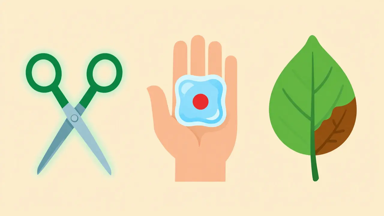
- Liver Transplantation: This is often the best option for patients with poor liver function. It removes both the cancer and the diseased liver. The Milan Criteria (one tumor ≤5 cm or up to three tumors ≤3 cm) generally determine eligibility.
- Surgical Resection: If your liver function is still good (Child-Pugh Class A) and the tumor is localized, surgeons can cut out the cancerous part while leaving the healthy liver intact.
- Ablation Therapy: For smaller tumors (<3 cm), doctors can destroy cancer cells using heat (radiofrequency ablation) or cold (cryoablation). This is less invasive than surgery and has high success rates for early-stage disease.
If the cancer is more advanced, treatments shift to controlling growth rather than curing. This includes transarterial chemoembolization (TACE), targeted therapies like sorafenib or lenvatinib, and immunotherapy combinations. The key takeaway? Early surveillance keeps you in the "curative" column, not the "management" column.
Barriers to Effective Surveillance
Knowing what to do and actually doing it are two different things. Studies show that only 30-50% of eligible cirrhotic patients in the United States receive guideline-concordant surveillance. Why the gap?
- Provider Awareness: Only 45% of hepatology practices have formal HCC surveillance pathways. Many primary care doctors simply don’t know when to refer.
- Patient Follow-Up: Between 25-40% of patients miss scheduled appointments. Fear, cost, or simple forgetfulness play a role.
- Disparities: Data reveals significant gaps. White patients are more likely to receive surveillance (52.3%) compared to Black patients (34.1%). Insurance status also matters, with privately insured patients seeing higher rates than those on Medicaid.
To fix this, successful programs use automated EHR reminders, patient navigators to reduce no-shows, and dedicated radiologists who specialize in liver imaging. If you’re a patient, advocate for yourself. Ask your doctor: “Am I being screened for liver cancer?”
Future of HCC Detection
The field is moving fast. By 2027, experts predict MRI might replace ultrasound for 30-40% of high-risk patients as costs drop. New biomarkers, like the GALAD score (combining gender, age, AFP-L3, AFP, and des-gamma-carboxy prothrombin), offer 85% sensitivity for early detection. AI-assisted tools, such as Medtronic’s LiverAssist, are already improving small lesion detection by nearly 20%. The goal remains the same: find it early, treat it effectively, and save lives.
Who needs HCC surveillance?
Anyone with cirrhosis, regardless of the cause (hepatitis, alcohol, NAFLD), should be considered for surveillance. Specifically, adults with Child-Turcotte-Pugh Class A and B cirrhosis are strong candidates. Those with Class C cirrhosis may be excluded unless they are on transplant waiting lists.
How often should I get an ultrasound?
Every six months. This interval is chosen because HCC tumors typically grow slowly enough that a 6-month check allows detection while the tumor is still small and treatable.
Is AFP testing necessary if I’m getting ultrasounds?
It is conditionally recommended. Ultrasound alone misses some tumors. Adding AFP blood tests can catch cancers that ultrasound overlooks, though it can also produce false positives. An AFP level >20 ng/mL requires immediate follow-up imaging.
What happens if my ultrasound shows a spot?
If a mass larger than 1 cm is found, you will undergo multiphase contrast imaging (CT or MRI). These scans provide detailed views of blood flow patterns, which help diagnose HCC without always needing a biopsy.
Can lifestyle changes prevent HCC in cirrhosis?
While lifestyle changes like stopping alcohol, managing weight, and treating viral hepatitis can slow liver damage, they do not eliminate HCC risk once cirrhosis is established. Surveillance remains critical even with healthy habits.