 Apr, 15 2026
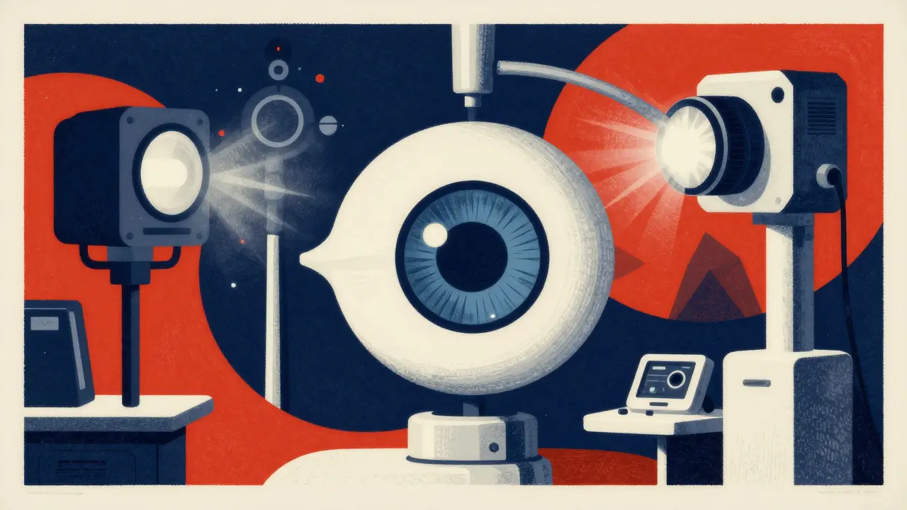
If you've ever been to an eye doctor and felt like you were stepping into a sci-fi movie with all the flashing lights and high-tech cameras, you're not alone. Modern eye care relies on a suite of imaging tools that let doctors see through the layers of your eye without actually touching them. Whether you're dealing with a sudden blur in your vision or managing a chronic condition, these tools are the difference between a "best guess" and a precise diagnosis. Let's break down how these technologies work and why your doctor might choose one over the other.
| Technology | What it Sees | Invasive? | Key Strength |
|---|---|---|---|
| Fundus Photography | Surface of the retina | No | Clear visual record/documentation |
| OCT | Cross-sectional layers | No | Extreme detail of retinal thickness |
| Fluorescein Angiography | Blood flow & leakage | Yes (IV Dye) | Detecting active vessel leaks |
| OCT Angiography (OCTA) | 3D blood vessels | No | Fast, dye-free vascular mapping |
The Basics: Fundus Photography
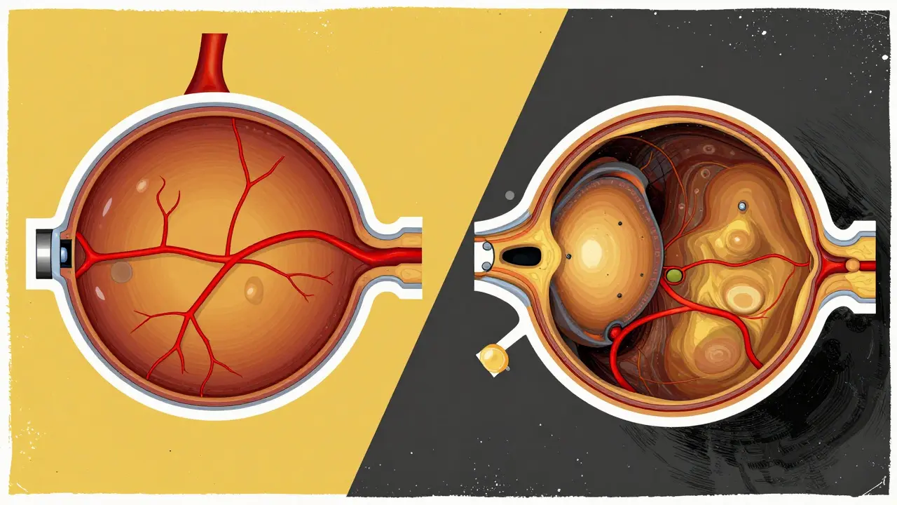
Think of Fundus Photography is a specialized digital photography technique used to capture high-resolution images of the interior surface of the eye as a high-definition "map" of your retina. It focuses on the posterior pole-the back part of the eye-including the macula, the optic disc, and the retinal blood vessels. Using cameras like the Zeiss FF 450+, doctors can spot obvious signs of damage, such as hemorrhages or exudates, which are essentially fatty deposits that leak from damaged vessels.
While these photos are great for documentation and tracking how a disease progresses over years, they only show the surface. It's like looking at a photo of a house from the street; you can see if the windows are broken, but you can't tell if there's a leak in the plumbing behind the walls. That's where more advanced imaging comes in.
Slicing Through the Eye: Optical Coherence Tomography (OCT)
If fundus photography is a 2D photo, Optical Coherence Tomography (or OCT) is like an ultrasound, but it uses light instead of sound. OCT is a non-contact imaging tool that produces high-resolution cross-sectional images of the retina, choroid, and optic nerve. By bouncing light waves off the different layers of the eye, it creates a detailed "slice" that allows doctors to see exactly where fluid is building up or where tissue is thinning.
There are two main types you might hear about. Spectral-Domain OCT (SD-OCT) became the gold standard in the late 2000s, offering an axial resolution of about 5-7μm. However, newer Swept-Source OCT (SS-OCT) can penetrate deeper into the eye, capturing up to 400,000 A-scans per second. This is crucial for seeing the deeper choroidal structures that SD-OCT might miss. For example, in cases of macular holes, OCT is the undisputed king because it reveals the actual physical gap in the retinal tissue.
Tracking Blood Flow: Fluorescein Angiography
When the problem isn't just the structure of the eye but how the blood is moving, doctors turn to Fluorescein Angiography (FA). This process is more invasive: a bright orange dye called fluorescein is injected into your arm. As the dye travels through your bloodstream and reaches the eye, a camera captures the flow in real-time. This technique, developed back in the 1960s, is still vital today because it shows leakage.
Why does leakage matter? In conditions like diabetic macular edema (DME), the blood vessels become "leaky," letting fluid seep into the retina. While OCT can see the fluid (the result), FA sees the leak (the cause). In a study of diabetic macular edema, FA actually showed higher sensitivity than SD-OCT for detecting these specific leaks. The downside? It takes 10 to 30 minutes, requires an IV, and carries a small risk of allergic reactions to the dye.
The New Frontier: OCT Angiography (OCTA)
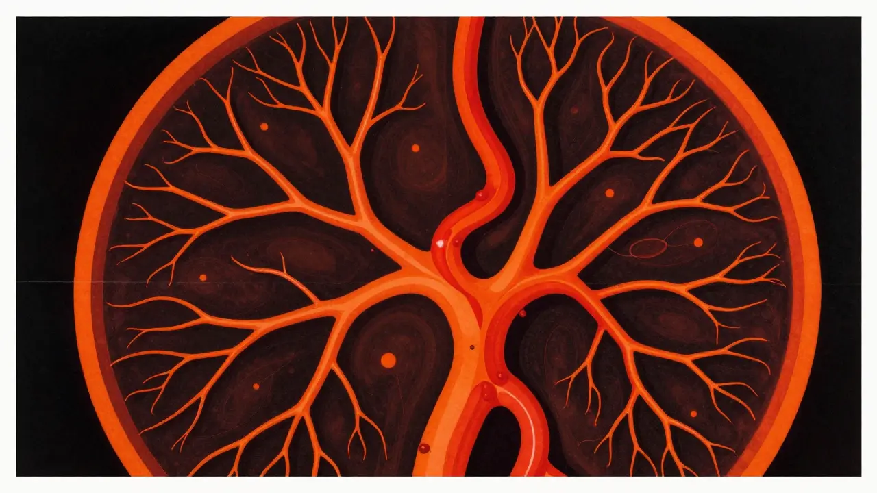
The game changed around 2014 with the introduction of OCT Angiography (OCTA). Imagine the detail of an OCT scan combined with the vascular mapping of an angiogram, but without the needles. OCTA is a non-invasive imaging method that uses the motion of red blood cells to create a 3D map of the retinal and choroidal blood vessels.
The speed is the biggest win here. What took 30 minutes with dye now takes seconds. It also lets doctors see the superficial, middle, and deep capillary plexuses without the "blur" caused by dye leakage. In cases of proliferative diabetic retinopathy, OCTA has proven superior in detecting neovessels-those fragile, new blood vessels that grow abnormally-especially around the optic disc. It's also a lifesaver for patients with rare conditions like Punctate Inner Choroidopathy (PIC), where it can find areas of non-perfusion (dead zones) that traditional imaging completely misses.
Choosing the Right Tool: The Multimodal Approach
You might wonder why doctors don't just use OCTA for everything since it's faster and safer. The truth is, no single tool provides the whole picture. This is why experts use a "multimodal" approach-combining several tests to cross-reference findings.
For instance, if a patient has Coats disease, a fundus photo might show yellow exudates. An OCT scan might then reveal that these exudates are actually sitting in multiple different retinal layers, along with small pockets of subretinal fluid. Finally, an FA might be used to pinpoint exactly where the abnormal vessels are leaking. Each tool fills a gap left by the other. While OCTA is great for mapping, it's still prone to "motion artifacts"-if you blink or move your eye during the scan, the image can blur, which is a common struggle with children or elderly patients.
| Clinical Goal | Preferred Tool | Why? |
|---|---|---|
| General Screening | Fundus Photography | Quick, visual record of the retina surface. |
| Retinal Thickness / Edema | OCT | Non-invasive cross-sections show fluid accumulation. |
| Vessel Leakage / Dye Flow | Fluorescein Angiography | Gold standard for seeing active leakage. |
| Vascular Mapping (Dye-Free) | OCTA | Fast, detailed 3D view of capillary networks. |
What to Expect During Your Appointment
Depending on the test, your experience will vary. For fundus photos and OCT, you'll likely just lean into a machine and look at a target light. Your pupils might be dilated with drops to give the camera a wider view. These tests are painless and quick.
If you're scheduled for a fluorescein angiography, it's a bit more involved. A nurse will start an IV, inject the dye, and the technician will take a series of photos over several minutes. You'll notice that your skin might take on a slightly yellowish tint for a few hours, and your urine will look bright yellow-this is normal as your body flushes out the dye.
With OCTA, the biggest challenge is staying still. Because the machine is mapping blood flow based on movement, any head shake or eye twitch can create an artifact that looks like a vascular abnormality but is actually just a "glitch" in the image. If you have trouble with fixation, your doctor might take multiple scans to ensure the data is clean.
Is OCT safer than Fluorescein Angiography?
Yes, generally. OCT is completely non-invasive and doesn't involve any dyes or needles. Fluorescein Angiography requires an intravenous injection of dye, which carries a small risk of allergic reactions or systemic side effects, making it a second-line choice if a non-invasive option like OCTA can provide the necessary information.
Can OCTA completely replace dye-based angiography?
Not yet. While OCTA is fantastic for seeing the structure of vessels and non-perfusion areas, it cannot detect "leakage." Since identifying where fluid is leaking from a vessel is critical for treating conditions like diabetic macular edema or retinal vein occlusions, Fluorescein Angiography remains essential for those specific diagnoses.
What is the difference between SD-OCT and SS-OCT?
The main difference is depth and speed. Spectral-Domain (SD-OCT) is the standard for most retinal scans. Swept-Source (SS-OCT) uses a different laser technology that allows it to penetrate deeper into the eye, providing much clearer images of the choroid (the layer beneath the retina), and it captures images much faster.
How long does it take to get these results?
For OCT, fundus photos, and OCTA, the images are available instantly on the screen. Your doctor can often interpret them during your appointment. Fluorescein angiography images are also captured in real-time, though the doctor may spend more time analyzing the sequence of dye flow before giving a final diagnosis.
Why do I need both an OCT and a Fundus photo?
They see different things. A fundus photo is like a map of the surface-it shows the layout of your retina and any obvious spots or bleeds. An OCT is like a cross-section-it shows the thickness and the layers. You need the map to know where to look, and the cross-section to see how deep the problem goes.
Next Steps and Troubleshooting
If you've been told you need these tests, don't panic. Most of them are routine and provide the data your doctor needs to start the right treatment. If you're feeling anxious about the IV for angiography, ask your doctor if OCTA is a viable alternative for your specific condition.
For those who struggle to keep their eyes still-such as young children or patients with advanced neurological conditions-let your clinic know in advance. They may be able to schedule your appointment when you're most alert or use specific stabilization techniques to reduce motion artifacts in your scans.
Jon lee
April 17, 2026 AT 02:28This is a really helpful breakdown for anyone feeling nervous about their next checkup. It's always easier when you actually understand what the machines are doing to your eyes!
Anna BB
April 17, 2026 AT 08:21It is just so fascinating how light can reveal the inner workings of our being... like a window into the soul, but for medicine!!! I love how these tools coexist to give a full picture...
Randall Barker
April 17, 2026 AT 22:07The obsession with 'faster and safer' is just a symptom of a society that refuses to endure any discomfort for the sake of truth. Fluorescein angiography isn't 'invasive' in any meaningful philosophical sense, yet people act like it's a root canal. We've become so soft that we value convenience over the gold standard of clinical evidence. It's a moral failure of the modern patient to demand a dye-free world when the dye is what actually shows the pathology. Why settle for a 3D map when you can see the actual biological failure in real-time? It's pathetic how we prioritize a few minutes of comfort over absolute diagnostic certainty. This push toward OCTA as a replacement is just lazy medicine catering to lazy patients. We should embrace the discomfort if it leads to a more honest diagnosis. Stop whining about needles and start caring about accuracy. The truth isn't always convenient, and medical imaging should reflect that reality. If you can't handle a tiny needle for the sake of your sight, you're just playing at being a patient. It's a joke.
Richard Moore
April 19, 2026 AT 13:20Whoa, let's keep it chill! 😅 I totally get the need for accuracy, but some people genuinely have phobias or allergies. OCTA is still a massive win for accessibility! 🚀
Heer Malhotra
April 20, 2026 AT 03:45The focus on Western medical standards here is quite presumptuous. India has developed world-class ophthalmic centers that utilize these exact technologies with far greater efficiency and volume than most US clinics ever could.
Autumn Bridwell
April 21, 2026 AT 18:53OMG I had this done last year and the technician was literally breathing down my neck the whole time!! I could feel his breath on my cheek and it was the most awkward experience of my entire life! I almost jumped out of the seat! 😱
Kim Hyunsoo
April 22, 2026 AT 02:48The way the light bounces around to create a slice is just... hypnotic 😵💫. It feels like some kind of digital alchemy happening inside the ocular globe (。◕‿◕。)
Theresa Griffin MEP
April 23, 2026 AT 13:36The clinical precision detailed here is exemplary. Efficient diagnostics save lives.
Dana Chichirita Nicoleta
April 24, 2026 AT 13:47I am absolutely overflowing with gratitude for this explanation! It is truly a beacon of clarity for those of us who navigate the terrifying waters of medical jargon without a map, and I simply cannot express how much I admire the way this simplifies such complex physics into something we can all grasp! My heart just leaps knowing that technology has advanced to the point where we can see the invisible currents of blood without always resorting to the needle, though the necessity of the dye is now perfectly clear to me! It is just such a magnificent era for human health!
Ben Ferguson
April 24, 2026 AT 21:35Speaking of the 'sci-fi' feeling, it reminds me so much of the way we describe the digital revolution in other parts of the world, where the leap from old analog systems to these high-speed scanners happened almost overnight in some regions, creating this surreal blend of 1960s clinics and 2024 lasers that you only see in the most eclectic parts of the global medical community, and it's just wild to think about the journey from simple photographs to these 3D capillary maps!
Josephine Wyburn
April 25, 2026 AT 13:42I'm just sitting here thinking about how stressed I get just thinking about the dye making my pee bright yellow 😭 like why does everything in medicine have to be so weird and gross? I can't even imagine the anxiety of lying there for 30 minutes while they take photos and I'm just worrying if I'm blinking too much or if the doctor is judging my retinal thickness 😩 it's honestly just too much for my nerves to handle ✨